Shoulder Rehabilitation Exercises From the FMS Library
Before we get into the exercises, it’s useful to understand a bit about the theoretical framework that underpins them.
An article by Brett Jones published at Functionalmovement.com highlights the rational – emphasised by Robert Burgess – that the the thoracic spine and ribcage are a movable spring.
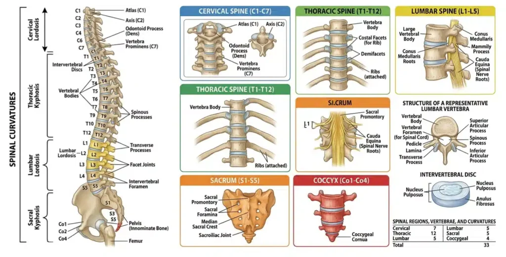
The graphic below provides a basic brake down of the spinal column.
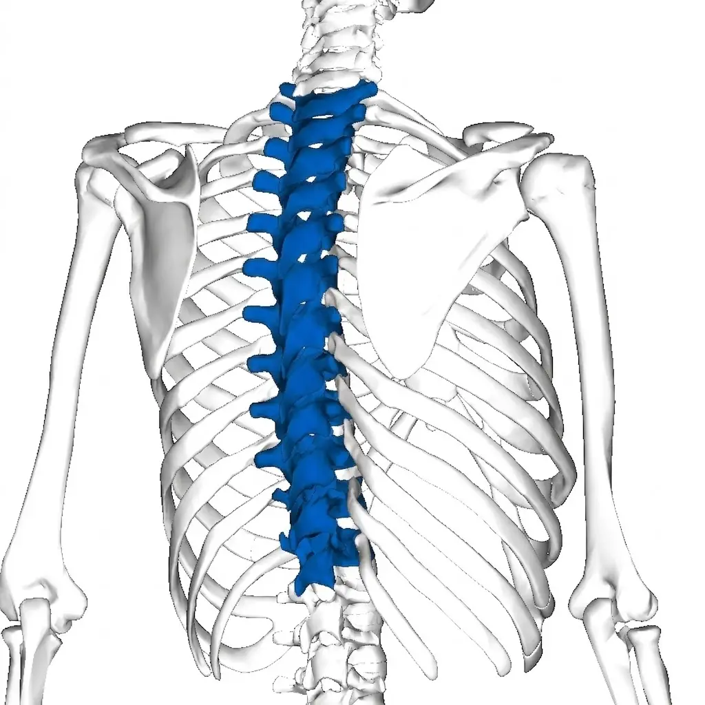
To clearly represent the thoracic spine you can refer to the blue segments in the following image.
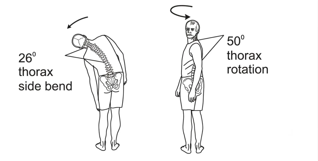
According to Buchalter et al 1988 and Willems et al 1996 the thoracic spine provides us with 50 degrees of rotation, 26 degrees of side bending, as pictured below.
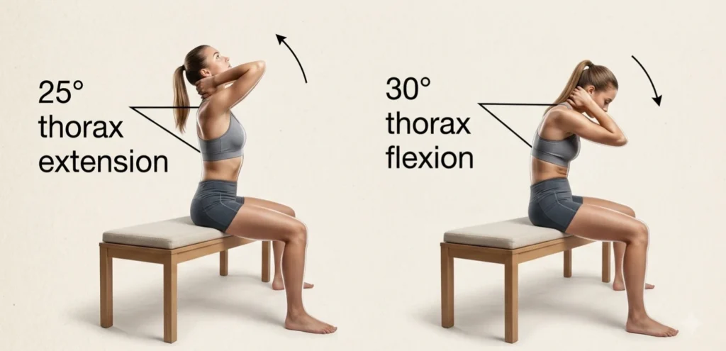
The thoracic spine also provides us with 25 degrees of extension and 30 degrees of flexion in sitting.
To emphasise their significance, Gracovetsky asserts “the pelvis and thorax rotate in opposing directions and together with side bending are the prime movers of human locomotion” (Gracovetsky, 1997).
Brett Jones asserts “The ribs connect up front on the sternum by way of a large series of cartilage joints – rather than being more solid “bone to bone” joints like the Sternoclavicular joint. These cartilage joints provide a more mobile situation and even the sternum has at least two semi-movable joints in its structure.”
As Brett emphasizes, if we are breathing correctly, using the diaphragm and intercostals (the muscles connecting your ribs) for respiration, there is significant movement in the thoracic spine and lower rib cage.
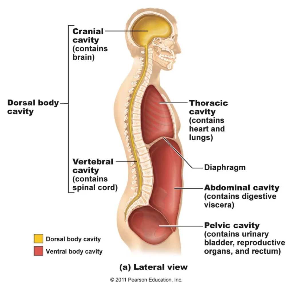
However, most people breathe into their upper chest which facilitates the development of excessive kyphosis (rounding of the upper back), tension in the anterior chain – across the front of the body – and a rigid rib cage.
Shoulder Mobility
By this point you might be wondering why we’ve spent so much time discussing the thoracic spine in relation to shoulder rehabilitation.
From a Functional Movement Screen (FMS) perspective, the relationship between the thorax (thoracic spine and rib cage) and the shoulder complex relies directly on the Joint-by-Joint Approach.
The thoracic spine is intended to be a mobile segment, while the joints of the shoulder – the scapulothoracic joint and glenohumeral joint – require that underlying mobility to achieve proper stability and positioning.
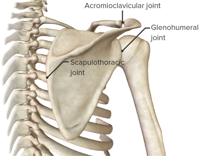
When thorax “opening” (thoracic extension and rotation) is limited, the entire kinetic chain of the shoulder suffers.
According to FMS literature, attempting to mobilize or strengthen the shoulder joint without first “opening” and unlocking the thoracic spine is treated as adding strength to dysfunction. The thoracic spine acts as the foundational dynamic anchor; if the anchor is locked, the arm cannot move cleanly.
Key Mechanical Benefits of Thoracic Mobility
1. Enables Full Scapular Upward Rotation & Posterior Tilt
The scapula sits directly on top of the posterior rib cage. For the shoulder to reach full overhead extension or bilateral internal/external rotation (as tested in the FMS Shoulder Mobility Screen), the scapula must tilt posteriorly and rotate upwardly.
Rounded Thoracic Spine (Flexion): Locks the scapula in protraction and anterior tilt, physically blocking full arm elevation.
Protraction: In the context of shoulder mechanics and thoracic mobility, scapular protraction refers to the shoulder blade sliding forward and outward around the rib cage—away from the spine toward the chest.
Anterior Tilt: In the context of scapular mechanics, scapular anterior tilt refers to the top of the shoulder blade (the acromion) tipping forward and down, while the bottom tip (inferior angle) lifts slightly off the rib cage.
Open Thorax (Extension): Restores the mechanical sliding surface for the scapula, allowing it to glide freely over the rib cage.
2. Protects the Subacromial Space
Thoracic extension directly influences subacromial clearance.
Subacromial clearance refers to the amount of space available in the subacromial space—the narrow gap beneath the arch of the acromion (the top bone of the shoulder blade) and above the head of the humerus (the upper arm bone).
This space is crucial because it houses important soft tissue structures:
- The Rotator Cuff Tendons (especially the supraspinatus)
- The Subacromial Bursa (a fluid-filled sac that reduces friction)
- The Long Head of the Biceps Tendon
When the thoracic spine remains flexed, arm elevation causes the head of the humerus to jam against the acromion process, causing subacromial impingement.
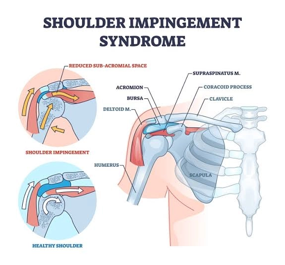
When you raise your arm overhead, adequate subacromial clearance is required so these soft tissues can glide freely without being pinched, compressed, or irritated against the hard acromion bone.
Opening the thorax lifts the acromion up and back, preserving the subacromial space for the rotator cuff tendons and bursa to clear safely during overhead movement.
3. Restores Symmetrical Rotational Mechanics
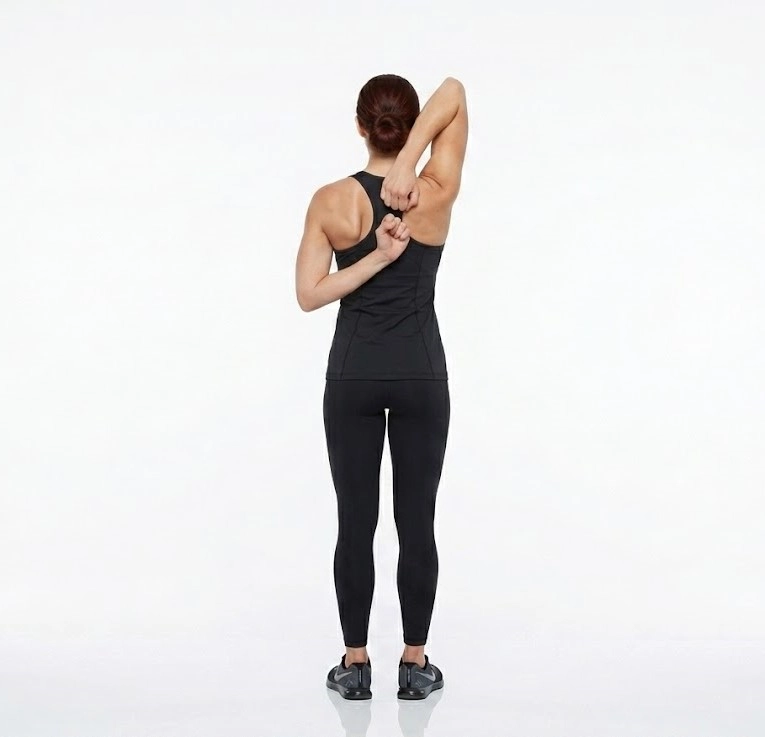
The FMS Shoulder Mobility assessment tests reciprocal pattern movement (one arm reaching overhead in external rotation, the other reaching behind the back in internal rotation). True shoulder reach requires thoracic rotation.
The Takeaway
If thoracic rotation is restricted, the body forces compensation by over-extending the lumbar spine or hyper-mobilizing the shoulder joint itself, leading to movement dysfunction and a lower FMS score (or pain clearing signals).
Simply put, you cannot fix the shoulder without first freeing the thorax. Attempting to build strength on top of a locked upper back only reinforces flawed mechanics and keeps the subacromial space compressed.
With the foundational anatomy and biomechanics established, we can now look at how the FMS translates these principles into targeted, practical movement.
The FMS Framework
The Functional Movement Systems (FMS) framework, provides corrective shoulder exercises designed to restore thoracic spine (upper back) extension, scapular stability, and shoulder mobility.
FMS follows a strict progression for shoulder correctives, starting with mobility work and advancing to loaded motor control.
1. Mobility & Soft Tissue (First Priority)
Before building shoulder strength, the thoracic spine and surrounding musculature must allow full range of motion.
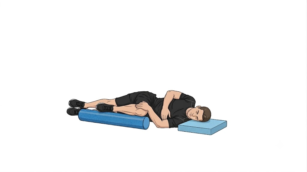
T-Spine Rotation with Rib Grab
The T-Spine Rotation with Rib Grab is a great breath assisted thorax opening drill. It uses deep diaphragmatic breathing to improve mobility. The Rib Grab is performed in a spine supported positions that creates minimal stability demands and lets the individual focus on the natural rotation and syncing the breath. Done with care and attention to the set-up it can lock out the lumbar and provide a great left to right appraisal of thorax rotation.
Setting Up: Start Position
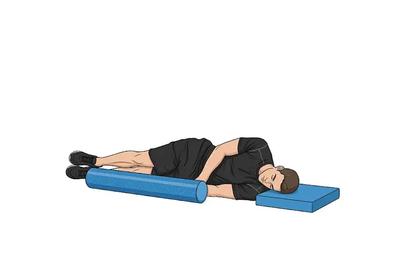
Lay on floor in a side lying position, flex the top hip to 90 degrees and support the knee with a foam roll and keep the foot on the ground. The head is supported by a towel roll. Reach under your ribs with the top hand. Begin rotating your top shoulder to the floor and pull the ribs in the direction you are rotating. Maintain contact between the knee and the foam roll. Then return to the starting position by rotating back to a neutral position.
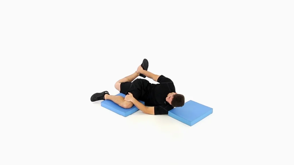
The Brettzel (Thoracic & Hip Mobility Integration)
The Brettzel takes thoracic rotation a step further by actively engaging the opposite-side hip’s anterior chain—testing hip extension and knee flexion simultaneously (for instance, rotating the shoulders to the right while stretching the left hip flexor and quad). This cross-body tension helps pinpoint whether restricted upper-back rotation is actually being anchored down by a tight hip or anterior chain. Just like the Rib Grab, this drill relies heavily on deep diaphragmatic breathing, with the goal of achieving that same smooth, uninhibited thoracic range.
2. Motor Control & Stability (Second Priority)
Once mobility is cleared, these drills teach the scapula to stabilize while the arm moves independently.
Quadruped Reach, Roll, and Lift: On hands and knees, reaching one arm forward, rolling the palm up, and lifting slightly to target the lower traps and serratus anterior.
Half-Kneeling Kettlebell Halo: Kneeling on one knee while slowly circling a kettlebell around your head. This forces core and shoulder blade stability under light changing loads.
Half-Kneeling Bottom-Up Kettlebell Hold: Holding a kettlebell upside-down by the handle. The instability forces deep shoulder stabilizers (rotator cuff) to fire automatically.
3. Pattern Integration & Loading (Final Step)
Once mobility and stability are established in static postures, dynamic movement is reintroduced.
Half-Kneeling / Standing Chops & Lifts: Using cables or bands across the body to integrate core rotation with diagonal upper-body pulling and pushing patterns.
Turkish Get-Up (Partial or Full): One of the ultimate FMS shoulder stability exercises, moving through multiple planes while maintaining a vertical arm position under load.
Single-Arm Farmer’s Carries: Walking with a heavy dumbbell or kettlebell in one hand to build reflexive shoulder packing and scapular depression.
Relationship with the Hip Joint
From a Functional Movement Screen (FMS) perspective, the relationship between the hip joint and shoulder mobility is governed by two foundational concepts pioneered by Gray Cook and Mike Boyle: Regional Interdependence and the Joint-by-Joint Approach.
Regional Interdependence states that seemingly unrelated, distant anatomical regions directly influence one another. Under the Joint-by-Joint framework, the human body alternates between mobile and stable segments:
FMS Joint-by-Joint Kinetic Chain
When the hip joint loses mobility, it triggers a cascade up the kinetic chain that directly compromises shoulder function. Here is how the hip joint impacts shoulder mobility through an FMS lens:
1. The Cross-Body Posterior Kinetic Chain (Anterior & Posterior Sling Systems)
The hips and shoulders are mechanically linked across the midline of the body through myofascial slings—specifically the Posterior Oblique Sling (connecting the gluteus maximus on one side to the contralateral latissimus dorsi via the thoracolumbar fascia).
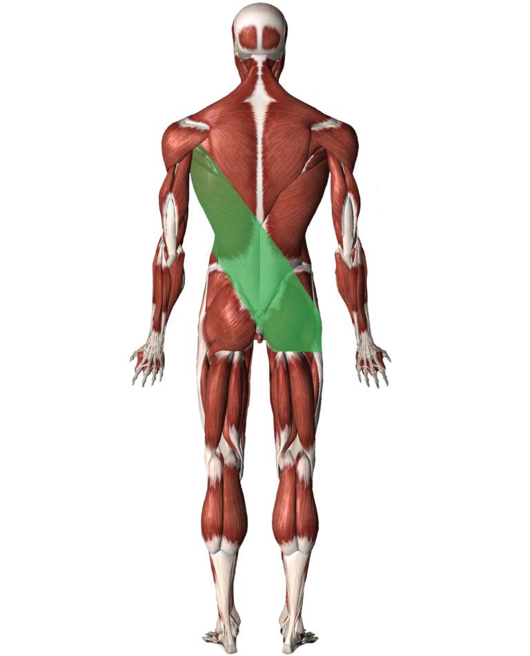
A myofascial sling is a continuous network of interconnected muscles, fascia (connective tissue), and ligaments that work together across joints to stabilize the skeleton and transfer force throughout the body during movement.
Rather than isolated muscles acting alone, myofascial slings act as dynamic tension cables. When one muscle in the chain contracts, it pulls through the fascia to activate and support other parts of the body along the same line.
- Hip Extension & Lat Tightness: If a hip lacks full extension, the latissimus dorsi on the opposite side often becomes chronically overactive or stiff to compensate for the lost stability/drive.
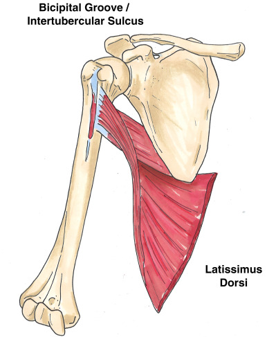
- Impact on the Shoulder: Because the latissimus dorsi inserts directly into the humerus and acts as a powerful internal rotator and depressor of the shoulder, an overactive lat locks the shoulder in internal rotation and restricts full overhead extension.
2. Preventing Lumbar Compensation
The lumbar spine is meant to serve as a stable base between the mobile hips and the mobile thoracic spine.
- When hip extension or hip rotation is restricted, the body forces range-of-motion from the nearest segment—the low back—causing excessive lumbar lordosis (arching).
- An arched lumbar spine forces the rib cage into an “open/flared” position, shifting the thoracic spine into extension and locking the scapula in an anteriorly tilted position.
- Outcome: The shoulder joint loses its solid, stable dynamic base, dramatically reducing reach and scoring lower on the FMS Shoulder Mobility screen.
3. High-Level FMS Pattern Interactions
The FMS never views shoulder mobility in isolated upper-body space; it evaluates how shoulder reach interacts with lower-body mechanics.
  [Restricted Hip Mobility]
             │
             ▼
  [Lumbar Hyper-Extension / Pelvic Tilt]
             │
             ▼
  [Rib Cage Flaring & Scapular Misalignment]
             │
             ▼
  [Compensated Shoulder Mobility Score]
The Active Straight Leg Raise (ASLR) Connection
In the FMS corrective hierarchy, the Active Straight Leg Raise (ASLR)—which measures hip mobility and core control—is prioritized high in the algorithm. If an individual has a baseline hip mobility deficit (scoring a 1 on ASLR), FMS principles dictate clearing or improving hip/pelvic control before trying to aggressive-stretch an asymmetrical shoulder. Restoring pelvis-on-hip alignment frequently frees up immediate overhead shoulder range without touching the shoulder joint itself.
The In-Line Lunge & Overhead Deep Squat
- In-Line Lunge: Requires opposite-arm/opposite-leg setup. Restricted hip flexors on the trailing leg pull the pelvis forward, making it impossible to keep the dowel aligned against the head, thoracic spine, and sacrum without shoulder strain.
- Overhead Deep Squat: Requires deep hip flexion combined with overhead shoulder extension. If the hips cannot sink into deep flexion, the center of mass shifts backward, forcing the shoulders to dump forward to prevent falling—a primary reason for a low score on the screen.
Key FMS Principle: The shoulder is often the victim, not the cause. Restoring hip mobility frees the pelvis, clears the lumbar spine to act as a stable anchor, and relaxes the broad back musculature so the shoulder can move cleanly.